Auerbach Plexus Is Present In
This collection of nerves is sandwiched between two layers of the muscularis externa the inner circular muscle. ICC were present in WWv mice at the deep muscular plexus in normal organization and numbers indicating that they are not dependent on the Kit protein and do not take part in generation of pacemaker activity.

Meissner S Plexus An Overview Sciencedirect Topics
The ENS operates both in conjunction with and independent of the peripheral nervous system.
Auerbach plexus is present in. However pathomorphological changes after. Ages 49 to 72 years. The most prominent symptom is constipation.
What kinds of tissue are present in the serosa of the GI tract. Nine patients 3 men and 6 women. Megacolon is most likely found in children that present with Down syndrome but another minor cause of megacolon is Chagas disease which can also cause megaesophagus.
Peristalsis - waves of smooth muscle contraction controlled by a nerve plexus called the myenteric Auerbachs plexus. Idiopathic esophageal achalasia is a motor disorder of unknown origin. Other symptoms may include vomiting abdominal pain diarrhea and slow growth.
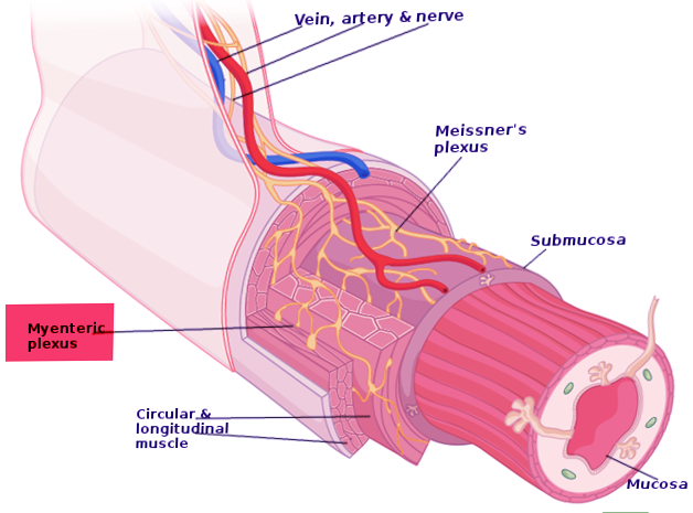
The submucosal plexus also known as Meissners plexus is situated in the submucosal region between the circular muscle and. The present study therefore was designed to extend our previous studies. Studies were performed on sera of 67 patients with primary achalasia of the esophagus.
The former requires general anesthesia and experience in interpretation 40. Is Meissner plexus present in Oesophagus. Hence no cell type had replaced ICC at their normal morphological position associated with Auerbachs plexus.
The absence of ganglion cells in the myenteric plexus Auerbach and the submucosal plexus Meissner of the colon over a variable length is the hallmark of Hirschsprung disease. Can the serosa and adventitia be present in the same area at the same time. Esophageal achalasia causes dysphagia following impaired relaxation of the lower esophageal sphincter due to the degeneration of Auerbachs plexus in the esophageal smooth muscle.
Produce distal intestinal segment that lack both meissner submucosal plexus and auerbach myenteric plexus. The neuronal loss observed in the disease has been suggested. Hirschsprungs disease HD or HSCR is a birth defect in which nerves are missing from parts of the intestine.
SummaryWe systematically studied the enteric nervous system of the alimentary tract in seven patients with Parkinsons disease. Complications may include enterocolitis megacolon bowel. Other articles where Auerbach plexus is discussed.
The choroid plexus is present in the ventricles of the brain. Nerve plexi exist within the bowel wall with Auerbachs plexus sandwiched between longitudinal and circular muscle layers and Meissners plexus located. We undertook a prospective study to determine whether microscopic changes were present in the myenteric plexus of patients with hypertensive lower esophageal sphincter nutcracker esophagus and diffuse esophageal spasm and if there was a correlation with lower esophageal sphincter pressure.
The myenteric plexus also known as Auerbachs plexus is located between the longitudinal and circular muscle layers of the esophagus stomach and small and large intestine. It is mainly made up of sensory nerves. In all patients characteristic inclusions histologically and ultrastructurally identical to Lewy bodies were found in Auerbachs and Meissners plexuses indicating that the plexus are also involved in Parkinsons disease.
Previous studies have often revealed an absence or reduction of ganglia in Auerbachs plexus in many patients with achalasia which has been postulated to be related to the elevated lower esophageal sphincter pressure in these patients. The submucous plexus as its name implies. Symptoms usually become apparent in the first two months of life.
The myenteric plexus is situated between the circular muscle layer and the longitudinal muscle layer in the lower esophagus stomach and intestines. Auerbach plexus is the interwoven network of the vagus nerve in the inner circular and outer longitudinal muscles of the muscularis layer of the visceral organs. We undertook a prospective study to determine whether microscopic changes were present in the myenteric plexus of patients with.
Auerbachs Plexus Throughout the gastrointestinal tract postganglionic parasympathetic neurons supply the glands and smooth muscle layers. Currently the leading theory states that Trypanosoma cruzi causes the destruction of Auerbachs plexus in the walls of the colon leading to the megacolon andor megaesophagus. The diagnosis can be confirmed by full-thickness rectal biopsy or suction rectal biopsy.
The cells of the Auerbachs myenteric plexus are located between the inner circular and outer longitudinal layers of the muscularis externa. We described recently the occurrence of autoimmune phenomena in patients with achalasia in particular circulating antibodies against Auerbachs plexus 1 2. Maqbool in Encyclopedia of Human Nutrition Third Edition 2013 The ENS and Gastrointestinal Motility.
Auerbach plexus named after Leopold Auerbach 1828-1897 also known by the name of myenteric plexus is a group of ganglia that run throughout the entire gastrointestinal tract and innervate its multiple layers of smooth muscle. Present in neonate with failure to pass meconium in the postnatal period followed by obstructive constipation. A causal relationship between eosinophil degranulation and.
So the correct answer is option B. Get FREE solutions to all questions from chapter DIGESTION AND ABSORPTION. The myenteric plexus Auerbachs plexus and the submucous plexus Meissners plexus.
The most prominent microscopical finding is a decrease in the number of ganglion cells in the Auerbach plexus. Recently peroral endoscopic myotomy POEM has become one of the preferred treatment options for esophageal achalasia. In doing so they coordinate peristalsis and digestion.
Cell from cecum to rectum is disrupted. Auerbachs plexus or myenteric plexus is a part of the enteric nervous system that located between the longitudinal and circular layers of muscularis externa in the gastrointestinal tract. Watch complete video answer for Auerbachs plexus is present in of Biology Class 11th.

Myenteric Auerbach Plexus Of The Colon Of The Present Case A B And Download Scientific Diagram

Stephen King 1974 Planning Cycle Jwt London Planning Cycle How To Plan Stephen King

A Schematic Representation Of The Gut Wall Myenteric Plexus Download Scientific Diagram

Enteric Nervous System Enteric Nervous System Nervous System Biochemical
Auerbach S Plexus


Pin By Apoorva Singh On Photographic Memory For Doctors Plexus Products Neurons Parasympathetic

Histological Findings In The Myenteric Plexus Of Colon A Download Scientific Diagram

Week 12 Gi Physiology Flashcards Quizlet
Auerbach S Plexus


Pin On Medicină

Pin On Histology Slides

Pin On Rad Tech

Solved In The Gastrointestinal Tract The Meissner S Plexus And The Auerbach S Plexus Occur Respectively In The

Stomach Plexus Products Flashcards Study Tools

Gi Tissue Ultra Structure Flashcards Quizlet

Pin On Histology Slides
Neuroanatomy Auerbach Plexus Article

Auerbach S Plexus Is Present In

Epithelial Cell Defense Mechanisms Of The Digestive System The Potential For Intraluminal Contents To Cause Sev Defense Mechanisms Digestive System Defense
Post a Comment for "Auerbach Plexus Is Present In"