Auerbach Plexus Histology
The ENS operates both in conjunction with and independent of the peripheral nervous system. Meissners plexus is located in the submucosa.

Gi Histological Anatomy Plexus Products Digestive System Physiology
For over 70 years our mission has been to provide educators with top-quality microscope slides for botany zoology histology embryology parasitology genetics and pathology.

Auerbach plexus histology. - myenteric auerbachs plexus - located between inner and outer layers of muscularis externa - submucosal meissners plexus - in submucosa. Study sets textbooks questions. Bellini Lorenzo 1643-1704 Italian anatomist who did some histology.
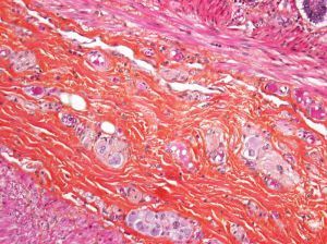
Histology for Pathology Gastrointestinal System and Exocrine Pancreas Theresa Kristopaitis MD. Auerbachs or the myenteric plexus is found between the outer longitudinal and inner circular muscle layers of the GI. In between circular and longitudinal muscle layers few myenteric or auerbachs plexus of nerve fibres are seenmiddle circular muscle layer is thicker.
The cells of the Auerbachs myenteric plexus are located between the inner circular and outer longitudinal layers of the muscularis externa. Auerbachs Plexus Throughout the gastrointestinal tract postganglionic parasympathetic neurons supply the glands and smooth muscle layers. Bertin Exupère Joseph 1712-1781 French anatomist who did some histology.
The plexus Auerbach consists of densely glyoxylic acid induced fluorescent GIF elongated ganglia with in general a longitudinal axis running parallel to the circular muscle layer and large dense interconnecting fibre tracts with primary secondary and tertiary subdivisions. Aging is believed to affect the structure and function of the enteric nervous system in the gastrointestinal tract. We offer an extensive collection of prepared slides for educators at all levels of instruction.
Only the latter two layers are seen here. In doing so they coordinate peristalsis and digestion. Maqbool in Encyclopedia of Human Nutrition Third Edition 2013 The ENS and Gastrointestinal Motility.
It has a dense network of veins rectal venous plexus and is thickened at the transverse folds. Each tissueorgan slide set has an explanatory accompanying text which desribes its structure function and role. Thirty male albino rats were used in this study and divided equally into three.
The muscularis externa consists of thick layers of smooth muscle. Auerbach Leopold 1828-1897 German anatomist who did some histology. Auerbachs plexus myenteric plexus Histology.
Renal columns of Bertin. Start studying Histology of the Upper GI Tract. The outer layer of the GI tract is either an adventitia or serosa.
Associated with myenteric Auerbachintramuscular plexus between circular and longitudinal muscle layers Have pacemaker function which facilitates active propagation of electrical events and mediates neurotransmission Have unique ultrastructure on EM with gap junctions between each other and smooth muscle cells. Auerbachs plexus lies between the layers of the muscularis externa throughout the tubular digestive system providing autonomic innervation to the smooth muscle of muscularis externa. It contains Auerbachs plexus.
The muscularis has the typical inner circular and outer longitudinal musculature between which the Auerbachs plexus lies. It contains ganglia from the parasympathetic system and nerve fibers from both the parasympathetic and. The submucosa contains loose connective tissue with blood vessels lymph follicles and the Meissners plexus.
The activating neurons use acetylcholine to stimulate the smooth muscles of the hollow organs while the. Made up of loose areolar connective tissuemeisseners plexus of nerve fibres are present. The submucosa is connective tissue.
The nerves in Meissners plexus. - histology of submandibular gland - mixed seromucous gland - SA serous protein-producing acini darkly stained and spherical in shape. Morphological Characterization of the Myenteric Plexus of the Ileum and Distal colon of Dogs Affected by Muscular Dystrophy Anat Rec Hoboken.
Auerbachs plexus located between circular and longitudinal muscle layers provides fibers from the autonomic nervous system to muscularis externa. Learn vocabulary terms and more with flashcards games and other study tools. Describe the components of the submucosal layer of the digestive organs Explain the location of Meissner plexus vs Auerbach plexus and describe the function of each Name the type of epithelium comprising the mucosa of the esophagus stomach small.
This section of dentaljuce has over 400 histological slides showing tissues from all organ systems in their healthy state. Meissners Plexus This section of the colon shows the cells of the Meissners submucosal plexus in close association with the smooth muscle of the muscularis mucosa. This work was designed to study the histological changes that might occur in the myenteric plexus of rat gastric fundus during aging.
Aim of the work. Nerve plexi exist within the bowel wall with Auerbachs plexus sandwiched between longitudinal and circular muscle layers and Meissners plexus located. The histology of the Auerbachs plexus is still not entirely known.
Colonic crypts lined by epithelial cells supported by the lamina propria are also visible. Auerbachs Myenteric Plexus-Located in bw outer circular layer and inner longitudinal layer. - Auerbachs plexus Muscularis externa of the stomach has inner oblique an inconsistent layer middle circular and outer longitudinal layers.
Consists of inner obliquemiddle circular and outer longitudinal muscle layers. Auerbachs Plexus Human section Microscope Slide Item 313798. The plexus comprises myenteric ganglia and the nerves emanating from them.
The plexus is a meshwork of unmyelinated axons and ganglia whose large neuronal cell bodies give rise to the postganglionic parasympathetic fibers in the plexus. Within it are lymphatic vessels and nerve plexuses. Auerbachs plexus epon 160x.
The myenteric neurons can be divided into activating cholinergic and inhibitory nitrogenergic neurons.

Choroid Histologia

Histology Of Gastrointestinal Tract Gastrointestinal Intestines Anatomy Plexus Products

Stomach Plexus Products Flashcards Study Tools

Auerbach S Plexus Plexus Products Microscopic Cells Auerbach

Digestive Anatomy In 2021 Digestive System Anatomy Medical Laboratory Science Anatomy
Plexus Myenteriques D Auerbach Avec Des Cellules Ganglionnaires Normaux Mo Plexus Myenteriques D Auerbach Avec Des Plexus Products Healthy Colon Auerbach

Digestive Tract Wall Plexus Products Science Illustration Digestion

Esophagus Histology Histology Slides Anatomy And Physiology Medical Studies
Pin By Hubert On Pathology Tissue Biology Anatomy And Physiology Study Of Tissues

Pin On Histology Slides

Dog Small Intestine Smooth Muscle Transverse Section 250x Smooth Muscle Mammals Muscular System Other System Muscle Plexus Products Muscular System

Auerbach Plexus Esophagus Histology Histo Love Plexus Products Histology Slides Med Student

Histology The Study Of The Microscopic Structure Of Tissues Histology Slides Microscopic Study

Histology Of Stomach Plexus Products Stomach Digestion

Intestine Plexus Plexus Products Lymphatic Anatomy And Physiology

Enteric Nervous System Enteric Nervous System Netter Medical Images Enteric Nervous System Plexus Products Brain Nervous System

Pin On Histology Slides

Pin By Brooke Bourgeois On School Stuff Digestive System Model Plexus Products Digestive System

Histology Of Esophagus The Esophagus Like Other Parts Of The Gastrointestinal Tract I Tissue Biology Loose Connective Tissue Stratified Squamous Epithelium
Post a Comment for "Auerbach Plexus Histology"